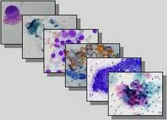
Symposium Breast Cytology
|
|
Symposium Breast Cytology |
SYMPOSIUM BREAST
CYTOLOGY 19. VISUALIZATION OF NONPALPABLE CHANGES IN BREAST AND TARGETED CYTOLOGICAL FINE-NEEDLE ASPIRATION Kardum-Skelin I1 , Šušterčić
D1 ,
Borovečki A1 , Parigros
K1 , Drinković I2 , Brkljačić
B2 , Brnić
Z2 ,
Viđak V2 , Kos
N3 Clinic of Internal Diseases and Department of Radiology KB
"Merkur"2, Zagreb; Private Clinic of Breast Diseases "Dr Nenad
Kos" Velika Gorica3 The golden standards in
breast diagnostics are: mammography, ultrasound and targeted FNA of changes
under the ultrasound control or stereotactically controlled FNA. The aim: 1. to analyse the technique of material
collection (FNA controlled blindely, by ultrasound or stereotactically); 2. to analyse the
number of FNAs performed in each patient and 3. to isolate the incidence of malignant
lesions smaller than 1 cm or incidence of
microcalcificate presence. The examinees and methods: for the last five years, in
3018 patients, 4264 FNAs by cytologists have been performed. In 4009 cases it was guided
by ultrasound, in 51 stereotactically guided, and in only 49 cases blindly
(palpationally). A fine-needle was used, and the slides were stained according to
Pappenheim (MGG). The results: 1. 1,4 FNAs on each patient; 2. blind FNAs
only in 1,1% cases; 3. malignant diagnosis determined in 237 FNAs (7,75%); 4. 17,2%
malignant lesions were smaller than 1 cm or only microcalcificates or in situ neoplasms were identified. With the introduction of targeted breast FNAs under the
ultrasound control or stereotactically with mammograph, it is possible to detect, early
and on time, a malignant growth of minute non-palpable lesions or even only changes with microcalcificates. SYMPOSIUM BREAST CYTOLOGY 20. ADVANTAGES AND VALIDITY OF CYTOLOGICAL FINE-NEEDLE ASPIRATION OF PALPABLE BREAST LESIONS GUIDED BY ULTRASOUND Knežević-Obad A, Dodig D Clinical Department of Nuclear Medicine, Rebro In a two-year period, 2.464 patients were examined by
ultrasound. Due to suspectable changes in the breast, 917 ultrasoundly controlled
cytological FNAs were performed. 820 cytological findings were in favour of benign breast
lesion without the need for further
therapeutical treatment. Fibroadenoma was diagnosed in 50 patients, and carcinoma
in 47 (later on histologically verified). 22 out of them were palpable, and, therefore,
besides a targeted cytological FNA ultrasoundly controlled, FNAs without the ultrasoud
control were performed in these cases, too. In
6 of them the adequate material for cytological analysis was not obtained by
"blind" FNA (malignant cells were not found). The incidence of desmoplasia,
tumorous necrosis, intense vascularisation, and tumor
in cystically changed breast were the causes of obtaining inadequate material for
cytological analysis. All the mentioned parametres can be removed if a
cytological FNA is performed under the ultrasound control, and with adequate blood suppy
in the suspected nodes by using colorDopler. Such method of obtaining material for a
cytological analysis ensures cytology an
adequate position in the diagnostic algorithm of breast examination, i.e. increases the
sensitivity and specifity of the method itself.
SYMPOSIUM BREAST CYTOLOGY 21. CYTOMORPHOLOGY OF ASPIRATED BREAST FRAGMENTS AFTER RADIATION - VALUE AND POSSIBLE ERRORS Mirjana Marković-Glamočak, Dubravka Boban,
Mirna Sučić, Šimun Križanac, Sunčica
Ries, Koraljka Gjadrov-Kuveždić Clinical Department of Laboratory Diagnostics, Department of Cytology, KBC Rebro We have analysed morphological
changes of glandular breast epithelial cells after the non-radical operation of breast
carcinoma and radiation. The aim of the work was to establish the possibilities of
cytological analysis of morphologically palpable changes. We have monitored 50 patients after the operation and
radiation, and analysed 71 aspirated fragments. In 12 patients the biopsy and pathohistological
verification have been carried out. In two cases carcinoma was cytologically diagnosed which
was further pathohistologically confirmed. In 3/10 (33.3%) cytologically suspected aspirated
fragments, recidivism was confirmed by a repeated FNA, and in 7/10 of suspected changes
only benign changes were revealed (2 mild ductal proliferation, 2 florid ductal
proliferation , 3 adenoses). In 16 patients the changes disappeared after the first
control and in 5 after the second control FNA. Cytodiagnostics is useful in evaluation and monitoring of
palpable changes in the breast after radiation,
but its role is limited. A cytologist has to know if the patient has ever been, and
exactly when, radiated. The test results of changed cells, after the period when these
changes were not present, suggest a disease recurrence. SYMPOSIUM BREAST CYTOLOGY 22. SIGNIFICANCE OF A TEAM APPROACH IN DIAGNOSTICS AND TREATMENT OF BREAST DISEASE Štoos-Veić T, Kaić G, Znidarčić Ž, Borković Z, Kozjak D, Stanec Z, Križanac Š Clinical Hospital "Dubrava" The development of medicine has provoked the need for
mutual expert work in different medical
professions in order to solve a great part of pathological
conditions. The manner of such cooperative work depends, certainly, on the type of a pathologic process, but the
results also depend on appropriate passing of information
by each participant, not only in his own branch, but also on the role of other expert he/she cooperates with. The presentation of our patient with both-sided breast
tumor shows the importance of adequately organised team approach in this problem area. The breast secretion is recognized as a significant
symptom of breast disease. The cytological examination is a part of initiative
diagnostical procedure, by which a greater secretive caused by benign changes in the
breast is revealed. Only in a smaller number
of cases, malignant tumor cells are found in secretion. There are cases in which this is the
only indication of breast malignoma that
cannot be confirmed by other diagnostical procedures. In our patients, the
diagnostical procedures were performed intentionally in order to detect both-sided tumors
whose only manifestation was a secretion from breasts with malignant tumor cells. If it
was not for the consistant cooperation among cytologists, radiologists, ultrasound
operators, surgeons and pathologists, this, nowdays, surgically curable malignant breast
tumor, could not be revealed in time. SYMPOSIUM BREAST CYTOLOGY 23. IMPORTANCE OF BREAST CYTODIAGNOSTICS IN A TEAM WORK Staklenac B, Hihlik-Babić
I, Margaretić D, Dmitrović B Clinical Hospital Osijek, Department of Clinical Cytology INTRODUCTION: Cytodiagnostics is a complementary method in multidisciplinary approach to breast diseases. AIM: The evaluation of cytodiagnostics by comparing the
mammographic, ultrasound and PHD results in 10 patients with breast nodes. MATERIAL AND METHODS: In the period between January 1, 1997 to March 3, 2000 in
the Cytological Unit of KB Osijek, 1591 breast FNAs were performed, out of which 174
(10,9%) under the ultrasound control. The material was obtained by fine needle aspiration of 22 G and 10 ccm syringe, then dried
in the air and stained by standard May-Grunwald Giemsa method. RESULTS: Out of the 1591 performed breast FNAs, 660 (41,48%) were
inadequate, 686 (43,12%) were benign, 113 (7,10%) suspectable and 132 (8,30%) malignant.
The mentioned methods were compared in 10 patients. The correspondence was revealed in 4
of them (regardless to whether it was benign
or malignant disease), while in 6 there was disagreement in some of them. CONCLUSION: The cytodiagnostics is a useful method whose aim is to
reduce the number of unnecessary operations, and, at the same time, to detect cancer in
the earliest possible stage because this makes patients´ survival longer. SYMPOSIUM BREAST CYTODIAGNOSTICS 24. CYTOLOGICAL PRESENTATION
OF PATHOLOGICAL CHANGES IN BREAST FROM TEN-YEAR WORK Gović A General Hospital Šibenik All types of pathological changes in the breast with a
special attention paid to the age structure in patients and their correspondence to
pathohistological results have been presented by retrospective analysis of cytological
results. 400 patients were examined in the monitored period. The number of examined patients is rising with the age
regardless to results of cytomorphological tests on changes. In the largest number of patients, regardless to the age
goups, benign changes with domination of fibrocystic disease were detected. The observations in work suggest a further detailed
examination, including cytological analysis in the monitoring of all pathologic changes in the breast. POSTER SECTION 25. PRESENTATION OF A PATIENT WITH NON-HODGKIN LYMPHOMA
AND BREAST CARCINOMA Sučić M1 , Boban
D1 ,
Marković-Glamočak M1 , Ries S1 , Gjadrov-Kuvedžić K1 , Podolski
P1 ,
Metelko-Kovačević J3 , Jakić-Razumović J4 , Hutinec
Z4 ,
Hlupić Lj4 , Budišić
Z4 1 Clinical Department of Laboratory Diagnostics, 2 Department of Oncology,
3Department of Hematology, 4Clinical Department of Pathology, KBC Zagreb The incidence of 2 or more malignant tumors can be
synchronic or metachronic. Simultaneous-synchronic proliferation of two malignant tumors
is more frequent in certain hereditary syndromes. Metachron-non-simultaneous occurance of
malignant tumors is connected to the influence of chemo-therapy and radiation which
damages cell deoxyribonucleic acid (DNA) and causes immunosurpression in patients. A patient, 66 -year old, is presented in the work, in
which, simultaneously, non-Hodgkin lymphoma
(NHL) and breast carcinoma were detected. Simultaneous incidence of breast carcinoma and
malignant hematologic disease is frequent in patients with Li-Fraumeni syndrome with the
hereditary mutation of p53 gene. The patient visited an oncologist because of the swelling on the left side of neck and a node in left breast. After the diagnostical processing, left breast carcinoma was detected in the patient. In the aspirated left breast fragment, malignant epithelial cells were found and immunocytochemically they were positive on cytokeratin. By clinical processing according to the TNM-classification of disease affection , the T2N×M0 stadium was determined. The cytomorphological findings in the aspirated fragment of the neck node suggested NHL of centro-blastic-centrocytic type. Llymphoma cells were LCA (leukocyte common antigen) positive as well as cytokeratin negative. NHL of tumorous cells, originating from follicle centre, was pathohistologically diagnosed in the patient´s neck node. The tumorous cells were LCA and CD20 positive, and EMA (epithelial membrane antigen), CD30 and cytokeratin negative. The identical lymphoma cells were detected in a bone-marrow biopsy, and by further hematological processing, NHL stadium IV was diagnosed in the patient. After a cytostatic treatment according to the CHOP scheme started, the regression of neck tumor occured and, after completing the first therapeutical part, a surgical treatment of breast carcinoma has been planned. |