Postgraduate Study
|
|
Postgraduate Study |
| DESCRIPTION OF PROFESSIONAL DEGREE The aim of the study is to introduce medical doctors, and in particular residents of clinical cytology with the foundations of this profession which has an exceptional interdisciplinary significance. Future clinical cytologists acquire specialised knowledge and skills during their residency in Clinical Cytology. The study programme is organised so as to offer information from fundamental sciences and applications of the most recent scientific methods in cytology, which, together with professional improvement, lays foundations for further research. The post-graduate professional study programme is
an obligatory part of the health specialization in Clinical Cytology (Regulations on
Resident Education of Health Providers of the Ministry for Health of the Republic of
Croatia, 1994) PROFESSIONAL DEGREE Master´s Degree ORGANIZER OF THE STUDY PROGRAMME University of Zagreb, Medical School DURATION OF THE STUDY PROGRAMME Four semesters CONDITIONS FOR ENROLMENT Graduates of the Faculty of Medicine who have passed the state professional exam; residents of clinical cytology have priority in enrolment.
LIST AND CONTENTS OF COURSES
THE PRINCIPLES OF MICROSCOPE TECHNIQUE Name of Head Nikola Ljubešić, M.D., Ph.D. Semester The first Number of Study Hours Lectures 10 (points: 1,0) Contents of Course Theoretical foundations of the light-microscopy. Magnifying glass and assembled microscope. The Abbe´s theory about the origin of picture in microscope. The power of differentiation and magnification. Practical handling with microscope. Phaso-contrasting, polarizational, fluorescent, laser "scanning" (LSM) and differentially interferential microscopes. A microscope with reflecting light and obscure range of sight. Theoretical foundations of electron microscopy (TEM, SEM, STEM). A short description of the basic preparative methods in electron microscopy. Geometrical measuring in microscopy. Microphotography, microprojection and usage of television and video equipment (VEC, AVEC). Foundations for image analysis. Literature Varićak B. Mikroskop: Teorijske osnove praktične mikroskopije. Zagreb (više raznih izdanja) Bradbury S. An Introduction to the Optical Microscopy. Revised Edition, Oxford University Press, 1989. Robards AW, Wilson AJ. Procedures in Electron Microscopy. John Wiley&Sons Ltd, 1993. Gerlach D. Das Lichtmikroskop, 2. uberarbeitete Auflage, Georg Thieme Verlag, Stuttgart, New York, 1985. Reimer L. Transmission Electron microscopy, 4th edition, Springer Verlag, Berlin, Heidelberg, 1997. Bancroft JD, Stevens A. Theory and Practice of Histological Techniques, 4th edition, Churcill Livingstone, New York, 1996. Manner of Examination Oral exam CELL BIOLOGY WITH ESSENTIAL HUMANE
GENETICS Name of Head Draško Šerman, M.D., Ph.D. Semester The first Number of Study Hours Lectures 16, seminars 4 (points: 2.0) Contents of Course Methods of cell examination. Cells of prokaryotae and eukaryotae. The information and energy flow in a cell. Molecular biology and organisation of genetic material. E u k a r y o t i c c e l l: The nucleus, chromatin, DNK, nucleolus, NOR, various RNK, histones. The structure and function of DNK: replication and activity of genes (copying), introns- exons, processing of pre-mRNK, hnRNK. The cell membrane system, cell membrane, nucleus integumentum. Endoplasmic reticulum, ribosomes, proteinic syntesis (transfer). Golgi´s complex, lysosomes, peroxisomes, cytoskeleton and cytosol: glycolysis, cytosol receptors, nucleocytoplasmic interactions. The role of steroid hormones, cytosol and nuclear receptors. Mitochondrion; the energy flow, Krebs´ cycle, oxidative phosphorilation, ATP, mitochondrial DNK and particularities of proteinic syntesis. Chloroplast, similarities and differences. P r o k a r y o t i c c e l l: The nucleotide, regulation of genetic activity: operon of lactose and histidine. Promoters, repressors, operators, terminators and atenuators, polycistronic mRNK. Escherichia coli and Salmonella typhimurium. Bacterial genetics and development of molecular genetics. The life cycle of a cell: interphase - division: mitosis, meiosis. Chromosomes (metaphasic, "lamp-brush" - and polyethene chromosomes). Reproductional biology, gametogenesis, insemination, growth, differentiation and morphogenesis, embryo, fetus, newborn. Transplatation of nuclei, genomic imprint, DNK methylation and regulation of transcription. Differentiation of erythrocytes: embryonal, fetal and grown-up hemoglobins, normal and abnormal hemoglobins. Mutations; in genes, number and structure of chromosomes. Mutagenesis, cancerogenesis and teratogenesis: ecotoxicology. Oxygen free radicals: inflammation, mutagenesis, cancerogenesis. F o u n d a t i o n s o f h u m a n e g e n e t i c s: Humane gentics, medical genetics and clinical genetics. Molecular biology in medical practice. One gene - one enzyme. Inborn metabolism abnormalities. Human caryotype, stripping of chromosomes, Q stripes - densitometry. The map of human genes: healthy and sick ones, X chromosome, Y chromosome. The map of human oncogenes and tumor of surpressing genes. The project of human genoma, trinucleotidyl recidives, length polymorphism of restrictional fragments, minisatellite DNK - fingers´ imprints. Fighting the cancer: American and European strategies and success/failure in the attempt to fight the cancer (proved treating methods versus preventions). Cytostatics as medicines with mutagenic and cancerogenic activitiy. Carcinogenesis is multigradual process; large intestine cancer. The interceptors of a carcinogene - desmutagenic. Your strategy of the cancer defence. Teaching Assistants D. Solter, M.D., Ph.D., V. Crnek, M.D., Ph.D., asst.prof., F. Bulić, M.D., Ph.D., asst.prof., H. Vlahović, M.D., Ph.D. M. Heffer-Lauc, M.D., Ph.D., asst.prof. Literature Šerman D. Mehanizmi genetske kontrole. U: Zergollern Lj, ur. Humana genetika. 3. izd. Zagreb: Medicinska naklada, 1994. Polšek D, Pavelić K (ur). Društveni značaj genske tehnologije. Institut društvenih znanosti "Ivo Pilar", Zagreb, 1999. Lewin B. Genes VII, Oxford University Press, Oxford, 1999. Solter D. Cloning and embryonic stem cells: a new era in human biology and medicine. Croatian MedJ 40: 309-318, 1999. Šerman D (ur.) Molekularna biologija (skripta). Zavod za biologiju Medicinskog fakulteta Sveučilišta u Zagrebu, Zagreb, 2000 Manner of Examination Oral exam. SELECTED CHAPTERS IN HISTOLOGY AND
EMBRYOLOGY Name of Head Ljerka Banek, M.D., Ph.D. Semester The first Number of Study Hours Lectures 16 (points 1,5) Contents of Course Histological structure of hematopoietic system and mesenchymal origin. Yolk-suck, blood islands, endothelium of blood vessels, embryonal liver, bone-marrow. The structure of lymph nodes, tonsils, spleen and thymus. General review of embryonal development of respiratory system .The structure of nose, pharynx, larynx, trachea, bronchi and lungs. The ultrastructure of lung alveoli. The blood-air barrier. Pleura. Histological structure of ovaries, oviduct, matrix, vagina, vulva The functional changes of these organs during generative female age and in the post-menopause. The development of lactic gland, changes in the structure of lactic gland during generative female age and in the menopause. The testis structure. Ductuli seminiferi. Sertoli and Leydig´s cells. Endocrine and paracrine regulation of spermatogenesis. Histological testis picture in different functional damages. General review of digestive tube with special attention paid to sub-microscopic structure of stomach, intestines, liver and pancreas. Embryonal development, structure and ultrastructure of thyroid gland. Histophysiology of thyroid gland in experimental environment. Co-assistants in Teaching Ž. Bradamante, M.D., Ph.D. Đ. Grbeša, M.D., Ph.D. R. Pezerović-Panijan, M.D., Ph.D. asst.prof., Lj. Kostović Knežević, M.D., Ph.D. Literature Fawcett DW. A Textbook of Histology. 12. izd. New York: Chapman&Hall, 1994. Sadler TW. Langman´s Medical Embryology. 7. izd. Baltimore: Williams and Wilkins, 1995. Selected articles of national and foreign authors. Junqueira LC, Carneiro J, Junqueira LC, Kelly RO. Basic Histology, 9th ed., Lange, 1998. Cormack DH. Clinically Integrated Histology, Lippincott Williams& Wilkins, 1998. Kerr JB Atlas of Functional Histology, Mosby, 1999. Burkitt HG. Wheater´s Functional Histology; A Text and Colour Atlas, Churcill livingstone, 2000. Moore KL., Persuad TVN. The Developing Human. Clinically Oriented Embriology, 6th ed., Saunders, 1998. Manner of Examination A written essay
SELECTED CHAPTERS IN PATHOLOGY Name of Head Šimun Križanac, M.D., Ph.D. Semester The first and second Number of Study Hours Lectures 34, exercises 12 (points 6,5) Contents of Course L u n g a n d m e d i a s t i n u m d i s e a s e s The morphological lung changes in AIDS, neoplasms of lung, pleura and mediastinum, neuroendocrine neoplasms. D i s e a s e s o f b l o o d a n d h e m a t o p o i e t i c o r g a n s Bone-marrow histology. Lymph node: non- malignant and malignant diseases. D i g e s t a l t r a c t d i s e a s e s Esophagus: Barrett´s esophagus, premalignant lesions, malignant lesions. Stomach: helicobacter pylori, chronic active gastritis, MALT - MALTOMI, intestinal metaplasia, possibility for differentiation of dysplasia from neoplasia in stomach mucus, ulcus disease, stomach neoplasm (expressions of oncogenic markers) Pancreas tumors (of benign, malignant structures in pancreas) Liver: iatrogenic lesions of liver parenchyma, chronic inflammatory liver diseases, virus markers and ISH virus, liver tumors. Intestines: polyp - sequence carcinoma (indication of disturbed oncogenic expression). P a t h o l o g y of f e m a l e g e n i t a l t r a c t Perineum, vulva, vagina: non-neoplasmic changes, condylomae, VIN, VAIN, invasive carcinoma - division and procedure. Uterine cervix: the inflammytory role of reserve cells and metaplasia in the growth of CIN, CIN, microinvasive carcinoma - division and procedure. Uterine body: functional endometria and hormonal changes of endometria. Metaplasia, hyperplasia and endometrial carcinoma. Neoplasms of mesenchymal origin. Division and procedure. Ovaries, oviduct and peritoneum: non-neoplasmic changes, primary and secondary ovarian neoplasms, neoplasmic markers. Division and procedure of ovarian neoplasms. Endosalpingiosis, endometriosis. Benign and malignant neoplasms of peritoneum and oviduct. The procedure. B r e a s t p a t h o l o g y The proliferative changes in breast, in situ carcinoma, invasive carcinoma, fibroadenoma and phyllode breast tumors. E n d o c r i n e g l a n d p a t h o l o g y Diffusive coloid thyroid gland struma. Nodose struma. Graves´ disease. Hashimoto and De Quervain thyreoditis. Benign and malignant neoplasms. P a t h o l o g y o f u r i n a r y t r a c t a n d m a l e g e n i t a l c e l l s: Iinflammations, benign and atypic hyperplasia and prostate neoplasms. Inflammations and neoplasms in seminals. Inflammations and neoplasms of kidneys and ureter. Inflammations and neoplasms of urinary bladder, urinary tube and male member. P a t h o l o g y o f l o c o m o t o r s y s t e m a n d s o f t t i s s u e s Neoplasms of bone-marrow and soft tissues. Neoplasms of non-explained histogenesis. DNA analysis, radiology. The role of biopsy in diagnostics and preoperational evaluation. The significance of immunohistochemistry as a diagnostical procedure. Teaching Assistants M. Belicza, M.D., Ph.D. S. Seiwerth, M.D.,Ph.D., asst.prof., S. Jukić,M.D.,Ph.D. D.Ljubanović,M.D., V.Šeparović,M.D.,Ph.D. M.Šćukanec-Špoljar, M.D.,Ph.D. J. Jakić-Razumović, M.D.,Ph.D., asst.prof. Literature Kurman RJ. Blaustein´s Pathology of the Female Genital Tract. New York - Berlin - Heidelberg: Springer-Verlag, 1994 GompleC, Silverberg SG. Pathology in Gynecology and Obstetrics. Philadelphia: JB Lippincott Company, 1994. Jukić S. and alt. Patologija ženskog spolnog sustava. Zagreb: AGM, 1999. Elaine S. Jaffe: Surgical Pathology of the Lymph Nodes and Related Organs; WB Saunder Comp, 1995 Dail DH, Hammar JP. Pulmonary Pathology. New York: Springer Verlag, 1997 Tavassoli SA. Pathology of the Breast, Appleton and Lange. Stanford, Connecticut, 1999 Damjanov I. Linder J. Anderson´s Pathology. 10 ed. Philadelphia: The CC Mosby company, 1995 Livosi VA. Surgical Pathology of the Thyroid. Philadelphia: WB Saunders Company, 1990 Ming SC. Pathology of the Gastrointestinal Tract; Williams and Wilkins 1998 Scheurer Pj. Liver Biopsy Interpretation. 3. ed. London: Bailler Tindall, 1997 Churg J. Sobin LH. Renal Disease. Classification and Atlas of Glomeular Diseases. New York: Igaku-Shoin, 1982 Churg J. Cotran RS, Sinniah R, Sahaguchi H, Sobin LH. Renal Disease. Classification and Atlas of tubulo-interstitial Diseases New York; Igaku-Shion, 1995 Churg J, Heptinstall RH, Olsen TS, Sobin LH. Renal Disease, New York: Igaku Shion, 1987 Cotran RS Kumar VK, Collins T, Robbins Sl. Pathologic basis of Disease. 6th ed. Philadelphia: WB Saunders Company, 1999 Enzinger FM. Weiss SV. Soft Tissue Tumors. 2.ed. Washington: Mosby company, 1994 Noltenius H. Human oncology. Pathology and Clinical Characteristics, 2.ed. Baltimore: Urban and Schwarzenberg, 1998 Magazines: - International Journal of Gynecological Pathology (New York) - International Journal of Gynecological Cancer (Cambridge) - Blood (Duluth) - Histopathology (Oxford) - Lung (New York) - Cancer (philadelphia) - Breast Cancer Research and Treatment (Hague) - Gastroenterology (Philadelphia) - American Journal of Surgical Pathology (New York) - Gut (London) Manner of Examination A written exam and identification of 10 histologic preparations after completing the course. ESSENTIAL
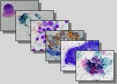
CLINICAL CYTOLOGY Name of Head Silvana Audy-Jurković, M.D., Ph.D. Semester The first Number of Study Hours Lectures 15, seminars 7 (points: 3,5) Contents of Course Introduction. The definition, historical development, indications, advantages, diagnostical value of clinical cytology, organisation of a cytological laboratory and control of working quality. Techniques of obtaining and processing biological material for cytological analysis. Fine-needle aspiration (of superficial organs and other superficial formations, palpable and non-palpable abdominal organs and formations, intrathoracal organs and formations, body cavities, bone-marrow); smear (mucus, skin); excretion (urine, sputum); imprint (of excised tissue); lavation (of body cavities, bronchi, bladder etc.). Appliances for collecting a specimen: aspirational needles, smear, spatula, brush etc. Equipment for targeted specimen collection: endoscopes, ultrasound, CT, EM. Types of specimens: aspirated fragments, smear, piece of excised tissue, excretion, washing. Preparation of specimens for cytologic analysis: type of preparation (smear, imprint, sediment, suspension); fixative type (for standard stainings according to Pappenheim - MGG and Papanicolaou, for different cytochemical and immunocytochemical stainings, for DNK cytometry and in situ hybridization); stainings (supravital, standard, cytochemical, immunological phenotyping. Analysis of processed material and procedures
used in clinical cytology. The approach to a cell analysis and cytologic smear
analysis. Non-stained, supravitally, standardly, cytochemically and immunocytochemically
stained smears. Digital processing of cytological
picture. Molecular diagnostics. Cytogenetics. Flow cytometry. Ways of expressing
cytologic results (description, cytologic diagnosis and classifications) and
interpretation. Clinico-laboratory correlation and procedure.Indications for application of particular diagnostical procedures. The completeness and competability of cytological and histological diagnostics. Rationalization of a diagnostic procedure. Efficiency of examinations in certain branches of clinical medicine. The role of team work in rendering the diagnosis. Teaching Assistants D. Batinić, M.D.,Ph.D., asst.prof. I. Črepinko, M.D., Ph.D. M. Halbauer, M.D., ph.D.,prim. V. Hitrec, M.D., Ph.D. I. Kardum-Skelin, M.D., M.S. V. Mahovlić, M.D., M.S. M.Marković-Glamočak, M.D., Ph.D. A.Ovanin-Rakić, M.D., M.S. Silvana Smojver, M.D., M.S. Trutin Ostović, M.D., M.S. A. Vince, M.D., Ph.D. Literature Bibbo M., ur. Comprehensive Cytopathology. Philadelphia: WB Saunders Company, 1997 Cardozo PL. Atlas of Clinical Cytopathology Weinheim: Edition Medizin, 1973 Koss LG. Diagnostic Cytology and Its Histopathologic Bases. 4 izd. Philadelphia: Lippincott Company, 1992. Črepinko I, Hauptmann E. Citodijagnostika. Medincinska enciklopedija, I. dopunski svezak. Zagreb: JLZ, 1976: 95-104 Ivić J. Citodijagnostika. Medicinska enciklopedija, I. dopunski svezak. Zagreb: JLZ, 1974: 104-7 Audy-Jurković S. Citološka klasifikacija cerviksa uterusa. Medicinska enciklopedija, II. dopunski svezak. Zagreb: JLZ, 1986:93 Ivić J, Audy-Jurković S. Citološka dijagnoza upala uzrokovanih mikroorganizmima onkogenog i teratogenog potencijala. Medicinska enciklopedija, II. dopunski svezak. Zagreb: JLZ, 1986: 93-94 Juretić A, Boban D, Čvorišćec D, Krajina Z, Labar B, Marković- Glamočak M, Sučić M. Citokemija i imunocitokemija u kliničkoj citologiji. Zagreb: Medicinski fakultet Sveučilišta u Zagrebu, 1992 Melamed MR, Lindmo Z, Mendelsohn ML. Flow Citometry and Sorting. 2. izd. New York: Wiley-Liss Inc. Boca Raton CRC Press, 1989 Baak JPA. Manual of Quantative Pathology in Cancer Diagnosis&Prognosis. Berlin: Springer Verlag, 1991: 382-95 Stavljenić-Rukavina A, Batinić D. Protočna citometrija u kliničko- laboratorijskoj dijagnostici. Klinički zavod za laboratorijsku dijagnostiku KBC zagreb, 1997 Labar B, Hauptmann E. Hematologija. Školska knjiga, Zagreb, 1999 Magazines: - Acta Cytologica (St. Louis) - Diagnostic Cytopathology (New York) - Analytical and Quantative Cytology (St. Louis) - Cancer (Philadelphia) - American Journal of Clinical Pathology (New York) - Applied Immunohistochemistry (Philadelphia) - Immunology (Oxford) - Cytometry (New York) Doctoral and master´s degree theses of national authors in the field of cytology. Proceedings of national and international conferences. Manner of Examination: Oral exam GYNECOLOGICAL CYTOLOGY Name of Head Silvana Audy-Jurković, M.D., Ph.D. Semester The first and second Number of Study Hours Lectures 39, seminars 23, exercises 8, (points: 11,0) Contents of course The development of gynecological cytology. Indications for cytological analyses. Advantages, diagnostical values and clinical usage of cytology. Specimen types, collecting and processing methods; the role of clinicians in determination of sensibility of cytologic analysis. The application of cytologic, cytochemical and immunocytochemical staining methods, as well as the methods of molecular biology in gynecological cytology. Cytomorphologics of normal cells of vulva, vagina, uterine cervix, endometryia, tube, ovaries A cytohormonic picture of vaginal epithelium from the fetal phase to post-menopause, including pregnancy, puerperium and lactation, and in endocrinologic disturbances and hormonal application. The cytohormonal changes on endometria). Cytology of amniotic liquid, determination of gestation period and early breaking of amnion. The tests for determination of X and Y chromosomes. Cytology of inflammation and inflammatory agents including determination of cleanness in a gynecological clinic. Benign proliferative changes. Epidemiology of cancer in female genital organs. The cytomorphological criteria in diagnosis and differential diagnosis of primary malignant tumors and their pre-stages, metastases and metastatic tumors. The significance of cytology in secondary prevention of malignant tumors, clinical graduation of malignant processes and determination of the surgery operation range. Cytological control of treated patients (surgery operation, radiation, chemotherapeutics, hormones). Clinical pictures, diagnostical methods and procedures, clinical classifications and treatment of diseases in the female genital tract. The classification of cytological findings. The role of clinical information in final cytological diagnosis and opinion. The team work with special stress on clinico-cytologic-histologic comparison. Teaching Assistants D.Buković, M.D.,Ph.D. A.Batinica-Grgurević, M.D.,M.S. I.Kuvačić, M.D.,Ph.D. N.Ljubojević, M.D.,Ph.D., asst.prof. V.Mahovlić, M.D.,M.S. B.Molnar-Stantić, M.D. A. Ovanin-Rakić, M.D.,M.S. M.Strnad,M.D., Ph.D., prim. V.Šimunić,M.D.,Ph.D., Đ.Šips, M.D. Literature Zajiček J. Aspiration Biopsy Cytology. Part2: Cytology of Infradiaphragmatic Organs. Basel: S.Karger, 1979: 38-103 Soost HJ, Baur S. Gynaekologische Zytodiagnostik. Stuttgart: Georg Thieme Verlag, 1990 Koss LG. Diagnostic Cytology and Its Histopathologic Bases. 4.izd. Philadelphia: J B Lippincott Company 1992: 251-686 Singer Z. Priručnik za ginekološku citologiju. 2.izd. Zagreb: vlast nakl, 1994 Ivić J, Audy-Jurković S. Vaginalna citologija u trudnoći. U: Dražančič i sur.. Porodništvo. Zagreb: Školska knjiga, 1994: 32-7 Ivić J, Audy-Jurković S. Citologija amnijske tekućine. U: Dražančić i sur. Porodništvo. Zagreb: Školska knjiga, 1994: 139-41 Audy-Jurković S, Ivić J. Citodijagnostika epitelnih atipija vrata maternica. U: Dražančić i sur. Porodništvo. zagreb: Školska knjiga, 1994: 388-92 Bibbo M, ur. Comprehensive Cytopathology. Philadelphia: WB Saundars Company, 1997: 93-323 Eljuga D, Dražančić A i sur. Prevencija i dijagnostika tumora ženskih spolnih organa. Zagreb: Nakladni zavod Globus, 1998 Magazines: - Gynecologia et perinatologia, Zagreb Selection from the literature listed in the course "Essential Clinical Cytology" Singer A, Monaghan JM. Lower Genital Tract Precancer. Colposcopy, Pathology and Treatment. 2nd ed. Blackwell Science Ltd, Oxford 2000 Manner of Examination Oral exam HEMATOLOGICAL CYTOLOGY Name of Head Željka Znidarčić, M.D., Ph.D., asst.prof. Semester The first and second Number of Study Hours Lectures 30, seminars 20, exercises 8, (points. 9,0) Contents of Course The historical development of cytology. Cytological significance and position in the routine and research work in hematology. The indications for cytological FNA of bone marrow, lymph node, spleen and liver. The methods of specimen collecting, its preparation and processing. The application of cytochemical, immunocytochemical, molecular and cytogenetical analyses in hematology. Normal development of hematopoietic cells (immunopoiesis, myelopoiesis). The structure, differentiation and maturation of lymphocytic system. The differentiation and maturation of myelopoiesis cells (erythropoiesis, granulopoiesis and thrombopoiesis). The cytological and phenotypic characteristics of normal hematopoietic cells in the maturation chain. Cell cultures in vitro, factors of growth. Morphological characteristics of non-malignant disturbances of cell myelopoiesis (anemia, anomalies and disturbances of granulocytes, monocytes and macrophages, disturbances in morphology and number of thrombocytes). Classification and morphological characteristics of stem cell diseases (aplastic anemia, isolated aplasia of the red lineage, myelodisplastic syndrome, chronic myeloproliferative syndrome). Non-malignant diseases of immunopoietic cells (morphologic and phenotypic particularities of lymphocyte and plasma cells in different compartments of lymphocytic system: peripheral blood, bone-marrow, lymph node). Neoplastic diseases of lymphocytic system: leukemic forms of chronic lymphoproliferative diseases, Hodgin and Non-Hodgkin malignant lymphomas, immunoproliferative diseases (morphologic, phenotypic, genotypic and cinetical features). Histiocytoses, parasites, "strange" cells (metastatic tumors) in bone- marrow. Acute leukemia (classification, morphology, cytochemical, immunophenotypic and genotypic characteristics). The spleen diseases. Morphological changes in bone-marrow and peripheral blood in patients with bone-marrow transplatation. The clinico-cytological correlation. Teaching Assistants D.Boban, M.D.,prim B.Jakšić, M.D.,Ph.D. I.Kardum-Skelin, M.D.,M.S. M.Sučić, M.D.,Ph.D. Literature Hauptmann E, Črepinko I. Osnove kliničke hematologije. Zagreb: Školska knjiga 1991 Jakšić B, Labar B, Grgičević D. Hematologija i transfuziologija. Zagreb: Jumena, 1998 Williams WJ. Hematology. 5.izd. New York: MC Graw-Hill, 1995 Hoffbrand AV, Petitt JE. Clinical Haematology - Sandoz Atlas. Basel: Sandoz LTD, 1988 Dilip KD. Lymph Nodes. U: Bibbo, ur. Comprehensive Cytopathology. Philadelphia: WB Saunders Company, 1991: 671-702 Lennert K. Histopathology of Non-Hodgkin Lymphoma. Feller AC, Berlin: Springer-Verlag, 1990 Labar B, Hauptmann E: Hematologija. Zagreb: Školska knjiga 1998 Eraldson RA: Diagnostic Transmission Electron Microscopy of Tumors. Raven Press New York 1994 Schumacher HR, Nand S. Myelodysplastic syndromes. New York&Tokyio: Igaku-Shoin, 1995 Harris NL, Jaffe ES, Diebold J, Flandrin G, Muller-Hermelink HK, Varidman J, Lister TA, Bloomfield CD. The World Health Organization classification of neoplastic diseases of the haemotopoetic and lymphoid tissues: report of the Clinical Advisory Committee Meeting, Airlie House, Virginia, November 1997. Histiopathology 2000; 36, 69-87 Magazines: - Blood (Duluth) - Haematology Pathology (New York) Selection from the literature listed in the "Essential Clinical Cytology" course Manner of Examination Oral exam. PULMONOLOGICAL CYTOLOGY Name of Head Mladen Pavlović, M.D., Ph.D. Semester The first and second Number of Study
Hours Lectures 16, seminars 20, exercises 4, (points 6,5) Contents of Course The role and value of cytodiagnostics in pulmonology. Types of material and collecting procedures. Material processing and specimen preparation. New methods and their application. Cytomorphology of respiratory system cells (upper and lower lung), pleura, other intrathoracic organs and tissues. The cytomorphological features of the respiratory system, pleura, mediastinum diseases. Atypia, proliferations, metaplasia and premalignant changes of respiratory system epithelium. The cytodiagnostics of pleural effusions. The cytomorphology of benign and malignant tumors of bronchi, lungs, pleura, mediastinum. The cytodiagnostics of secondary tumors of the same localizations. Intraoperational cytodiagnostics. Cytodiagnostics in controlling therapeutical effects. Posttherapeutical changes (irradiation, cytostatics) in normal and tumorous cells. Cytological findings (false positive and false negative results). The clinico-laboratory and cyto-histological correlation. Teaching Assistants M.Roglić, M.D., prim S.Smojver-Ježek, M.D., M.S. Literature Johnston WW. Respiratory cytology. U: Bibbo, ur. Comprehensive Cytopathology. Philadelphia:WB Saunders Company,1991:320-98 Naib ZM: Cytopathology. Boston, Little, Brawn and Co, 1996 Young JA. Colour Atlas of Pulmonary Cytopathology. New York: Harvey Miller Oxford University Press, 1985. Selection from the literature listed in the "Essential Clinical Cytology" course. Manner of Examination Oral exam. "Slide-test" ENDOCRINOLOGICAL CYTOLOGY
Name of Head Zdenko Škrabalo, M.D., Ph.D. Semester The second Number of Study Hours Lectures 16, seminars 12, exercises 4, (points: 5,0) Contents of course BREAST CYTODIAGNOSTICS The incidence of fibrocystic breast changes and breast carcinoma, and their correlation. The risk factors. The history of breast FNA. The possible damaging effect of the breast FNA. E x f o l i a t i v e b r e a s t c y t o d i a g n o s t i c s: The material collection for analysis, (secretion, scarificate), macroscopic and microscopic secretion appearance. Subareolar abscess, inflammation of Montgomery´s gland, the appearance of mammilla in eczema, and M.Paget. A s p i r a t i o n a l b r e a s t d i a g n o s t i c s: The indication and mode of FNA without and under the ultrasound control. Morphological picture of breast tissue, inflammatory changes, necrosis of fat tissue, fibrocystic changes with and without proliferation and atypia, fibroadenoma, macrocysts and carcinoma. The analysis and interpretations of breast nodes after non-radical surgeries (radiated benign and malignant cells). Tumor markers determined in the serum and/or aspirated breast fragments by immunocytochemistry or by techniques of molecular biology. Morphological changes in the breast during puberty, in adolescents and young women, in pregnancy, and in complementary hormonal treatment. Metastases into the breast. Metastases of the breast carcinoma into lymph nodes, skin, bone-marrow and other organs. The breast diseases in male-gynecomastia and carcinoma. Team work for breast diseases. The comparison of cytologic and pathohistological findings. CYTODIAGNOSTICS OF THYROID AND PARATHYROID The function of thyroid gland, paraphysiological bases of disturbances, diagnostical procedures of detecting pathological processes. The role of clinical cytology in diagnostics of thyroid gland diseases. The techniques of performing the thyroid FNAs without and under the ultrasound control. Cytological parametres of unchanged thyroid gland, inflammations and tumors (adenoma, Huerthle´s tumors, differentiated, non-differentiated and anaplastic carcinomas, medullary carcinomas, mixed thyroid tumors and lymphomas, metastasis of malignant tumors into thyroid gland and metastasis of thyroid carcinoma into lymph nodes). The usage of computerized morphometric analysis of a cell picture. The clinico-laboratory correlation of thyroid disease diagnostics. The parathormone and basic ideas about functional state of parathyroid glands. The role of clinical cytology and other diagnostical techniques in detection of localization, size and pathologic changes of parathyroid glands. The parathyroid FNAs controlled by ultrasound. Material processing (standard, cytochemical and immunocytochemical staining methods and computerized morphometric analysis of a cell picture). Interpretation of cytological findings of aspirated parathyroid tissues. The quantitative and qualitative changes of cytological parametres in adenoma, hyperplasia and carcinoma of parathyroid glands. The clinico-laboratory correlation. CYTODIAGNOSTICS OF TESTICLES AND EJACULATE Differentiation and development of the male reproductional system. The function of testicles in adulthood (spermatogenesis, spermiogenesis and hormonogenesis). The disturbances in functions and infertility in males. Diagnostical methods in processing the infertility (cytomorphology, cytogenetics, hormones, immunology, biochemistry and microbiology). Therapeutical procedures (pharmacotherapy, surgery, medically assisted inseminations, intracorporal and extracorporal). Cytomorphological analysis of ejaculate and urethra. The macroscopic (appearance, colour, consistency, volume and pH) and microscopic examinations (concetration and number of sperms, vitality, flexibility, HOS test, sperm morphology, other cellular elements). CASA techniques (computer assisted sperm analysis). The criteria of the World Health Organization. The techniques of isolation of kinetically and morphologically better sperms (density gradients and swim-up). Testicle cytology in examinations of male infertility. Material obtaining techniques. The cytological picture of testicles in oligozoospermia and in azoospermia (secretory and excretory). Testicle inflammations and tumors in cytological slides. Clinico-laboratory correlations in examination and treatment of male infertility. Teaching Assistants P. Cvitković, M.D., M.Halbauer, M.D., Ph.D., prim M.Marković-Glamočak, M.D.,Ph.D. I.Kardum-Skelin, M.D., M.S. P. Romac, B.S. in Biology., M.S. Literature Silverman JF.Breast, U: Bibbo M (ur), Comprehensive Cytopathology, Philadelphia: WB Saunders Company, 1997: 731-780 Zajiček J. Breast. U: Zajiček J. Aspiration Biopsy Cytology, part I. Cytology Supradiafragmatic of Organs, Basel: S.Karger, 1974: 136-94 Bibbo M.Abati A. The uniform approach to breast fine-needle aspiration Acta Cytol 1996; 40: 1119-1126 Sinham SK, Singh uR, Bhatia A. C-erb B2 oncoprotein expression. Correlation with Ki-67 labeling index and AgNOR counts in breast carcinoma on fine needle aspiration cytology, Acta Cytol. 1996; 40: 1216-1220 Albert U, Duda V, Hađi P. Imprint cytology of core needle biopsy specimens of breast lesions: a rapid approach to detecting malignancies with comparison of cytologic histopathologic analyses of 173 cases, Acta Cytol. 2000;44: 57-62 Droesse M. Aspirationzytologie der Schilddruese, Stuttgart: Schattauer, 1989 Eraldson RA. Thyroid Gland Neoplasms, U: RA Eraldson. Diagnostic Transmission Electron Mycroscopy of Tumors, new York: Raven Press, 1994: 797-801 Galera-Davidson H. Gonzalez-Campora R. Thyroid. U: Bibbo M(ur), Comprehensive Cytopathology, Philadelphia: WB Saunders Company, 1997: 673-701 Naib Z.M. The Thyroid Gland, U: Zuher M.Naib, Cytopathology, Boston -London: Little, Brown and Company, 1995: 517-535 Hargreave TB (ur), Male Infertility, London: Springer Verlag, 1994 Dieckmann KP, Skakkebaek NE. Carcinoma in situ of testis: review of biological and clinical features. Int.J Cancer, 1999; 83: 815-22 Cheville JC. Classification and pathology of testicular germ cell and sex cord-stromal tumors, Urol Clin North Amer, 1999; 26: 595-609 Papić Ž, Čolak B. Škrabalo Z. Fine needle aspiration biopsy (FNAB) of testicles, Diab Croat 1999; 28: 113-121 Cvitković P, Čolak B, i sur. Pristup neplodnosti u muškarca, Medicus 1999; 8: 193-206 Suggested magazines: - Thyroid (New York) ISSN 1050-7256 - European Urology, Basel, ISSN 0302-2338 -Fertility and Sterility Birmingam, Alabama ISSN 0015-0282 -Breast, London ISSN 09609776 Selection from the literature listed in the "Essential clinical cytology" course Manner of Examination Oral exam. GASTROENTEROLOGICAL CYTOLOGY Name of Head Branko Papa, M.D., Ph.D. Semester The second
Number of Hours Lectures 10, seminars 7, exercises 3, (points:4,0) Contents of course The cytological diagnostics value in gastroenterology. Types of specimen (aspirated fragment, smear, washing) and the techniques for obtainig material (targeted by endoscope, ultrasound, CT and EM). The value of complementary diagnostical procedures in gastroenterological diagnostics: cytochemistry, immunocytochemistry, kinetics and molecular biology. Cytomorphology of esophagus epithelial cells. Inflammatory changes (possibility of morphologic identification of specific agents). Barret´s esophagus. Premalignant lesions and tumors. Cytomorphology of stomach cells. Inflammatory changes in cells and possibilities of morphologic identification of agents - Helicobacter pylori. Atrofic gastritis, intestinal metaplasia, dysplastic changes. Cytomorphological criteria of premalignant and malignant epithelial lesions. Malignant lymphomas - MALTOMI. Normal cytomorphology of colon mucus. Inflammatory changes in cells - of bacterial, virus, viral and parasitic etiology and in chronic idiopathic intestinal diseases. Malignant diseases of small and large intestine (carcinoma, lymphomas, enterochromaphilic tumors). Cytomorphological characteristics of normal liver cells. Cytomorphology of inflammatory and chronic-degenerative liver changes. Primary and metastatic tumors. Cytomorphology of normal gall-bladder cells, inflammatory and malignant lesions. Cytomorphology of exocrine and endocrine parts of pancreas cells. Inflammatory changes (acute and chronic). Pseudocystic lesions. Benign and malignant pancreas tumors (cystadenomas, tumors of endocrine part, carcinomas). The clinico-cytological correlation. The competitiveness of cytological and histological findings in gastroenterological cytology. Teaching Assistants M.Barišić, M.D. T.Jeren, M.D., Ph.D. I.Kardum-Skelin, M.D., M.S. M.Katičić, M.D., Ph.D. S.Smojver-Ježek,M.D., M.S. Literature Geisinger KR. Alimentary Tract. U: Bibbo M,ur. Comprehensive Cytopathology. Philadelphia: WB Saunders Company, 1997: 413-44 Tao Liang-Che. Liver and Pancreas. U: Bibbo M, ur. Comprehensive Cytopathology. Philadelphia: WB Saunders Company, 1997: 827-864 Zuher MN. The Digestive Tract. U: Cytopathology. Little, Brown and Company, 1995: 311-348 Zuher MN. The Pancreas. U: Cytopathology. Little, Brown and Company 1995: 363-376 Zuher MN. The Liver. U: Cytopathology. Little, Brown and Company, 1995: 376-394 Erlandson RA. Diagnostic Transmission Electron Microscopy of Tumors. New York: Raven Press, 1994 Katičić M, Presečki V: Helicobacter pylori Izazov za medicinu. Zagreb: MGC Klovićevi Dvori, 1996 Magazines: - Gastroenterology (Philadelphia) Selection from the literature listed in the "Essential Clinical Cytology"course Manner of Examination Oral exam. UROLOGICAL CYTOLOGY Name of Head Željka Znidarčić, M.D., Ph.D. Semester The second Number of Study Hours Lectures 10, seminars 3, exercises 3 (points: 3,5) Contents of course The possibilities of cytodiagnostical application in urological practice. A cytological urinary examination - indications, technical preparation of urine. The urothelial structure and the appearance of urothelial cells in urinary sediment. The benign urotract diseases - kidney cysts, stones, inflammations (acute, subacute, chronic, specific-bacterial, viral, malacoplakia). Hematuria - the appearance of erythrocytes in urine sediment. Urothelial atypia - cytological characteristics and significance in diagnostics. Premalignant urothelial conditions - dyskaryosis. CIS, urothelial carcinoma. The characteristics of papillary urothelial tumors. Other urotract tumors - squamous, adenocarcinoma. The appearance of tubular kidney epithelium in urinary sediment. Renal tumors - classification and cytological characteristics, renal FNA techniques. Other materials for cytological researches - urethra secretion, separative urine from urether, washing of urinary bladder. Other techniques for urinary examination - flow citometry, genetic examinations. Inflammatory pathohistology, benign and atipic hyperplasia (stages). Prostate cancer - classification, differentional diagnosis of atypic hyperplasia and highly differentiated carcinoma. Other prostate tumors. The role of cytochemistry and immunocytochemistry in prostate disease diagnostics - tumor markers. Morphometric examinations. Teaching Assistants I. Kraljić, M.D., Ph.D., asst.prof. K.Trutin Ostović, M.D.,M.S. Literature Kern WH. Urinary Cytology. U: Bibbo M, ur. Comprehensive Cytopathology. Philadelphia: WB: Saunders Company (sec.ed.) 1997: 445-76 Shoog L, Myk-Tani E, Loewhagen T. Prostate. U: Bibbo M, ur. Comprehensive Cytopathology. Philadelphia: WB Saunders Company, 1991: 806-21 Katz RL. Kidneys. U. Bibbo M, ur. Comprehensive Cytology. Philadelphia: WB Saunders Company (sec.ed) 1997: 781-826 Kline TS. Handbook of Fine Needle Aspiration Cytology. 2.izd. Edinburgh: Churchill Livingstone, 1988: 365-92 Rathert P, Roth S, Soloway MS. Urinary Cytology. 2. izd. Heidelberg: Springer Verlag, 1991 Novak R, Tucak A i sur. Urološka onkologiija. Zagreb: Medicinska naklada, 1994 Kocjan GIL. Atlas of Diagnostic Cytopathology (sec.ed) 1997: 207-24 Magazines: -Journal of Urology (Baltimore) Selection from the literature listed in the "Essential Clinical Cytology" chapter. Manner of Examiantion Oral exam. SELECTED CHAPTERS IN CLINICAL CYTOLOGY Name of Head Tatjana Jeren, M.D., Ph.D. Semester The second Number of Study Hours Lectures 12, seminars 12 (points: 4,5) Contents of Course CYTODIAGNOSTICS OF HEAD AND NECK The historical review. Cytology in rhinology: Cytomorphologics of nasal mucus, aspirated sinus fragment, inflammatory diseases and tumors. Nasopharyngeal carcinoma. Immunocytochemical analysis. Cytology in otology: benign and malignant tumors. The salivary gland: inflammation, cysts and benign tumors, malignant and metastatic tumors. Neck cysts. Reactive lymphodenopathies. Metastatic neck tumors, analysis of glomus tumors, neurinoma, neurofibroma. CYTODIAGNOSTICS OF INFLAMMATORY AND DEGENERATIVE DISEASES OF LOCOMOTOR SYSTEM AND SOFT TISSUES cytomorphology of articular liquid. The modern classification, histogenesis, therapy and prognosis of mesenchymal tumors: malignant and benign tumors of soft tissues and bones. Cytochemistry, immunocytochemistry, flow citometry, cytogenetics as auxiliary diagnostical methods. CYTODIAGNOSTICS IN CHILDHOOD The particularities of cytodiagnostics in children. The bone marrow FNA adjusted to certain age. The method of collecting the peripheral blood smear in infants. Fetal hematopoiesis. Physiological differences of hematological findings in children. The blood cell anomalies pronaunced in children: the morphological erythrocyte analysis related to early detection of hereditary anemia, anomalies of the granulocyte linkeage. Morphological thrombocytic changes in peripheral blood. Tezaurismosis. Histiocytosis. Parasitosis. Tumors in children. Leukemia, myelodysplasia and malignant lymphomas in children. CYTODIAGNOSTICS IN SKIN DISEASES Some viral diseases of skin and visible mucus (h.simplex, h.zoster, varicella), intraepidermal achantolytic bullous dermatoses (the pemphigus group), subepidermal bullous dermatoses (the bullous pemphigoid group of herpetiform dermatitis). CYTODIAGNOSTICS OF CENTRAL NERVOUS SYSTEM
The liquor anatomy and physiology in different life ages. The techniques of liquor obtaining and preparation for analysis. Cytological findings in different inflammatory, degenerative, immunologic processes, bleedings, primary and secondary tumors. Immunocytochemistry. The application of PCR in liquor. The interpretation of cytological results. A differential diagnosis. Teaching Assistants I.Dobrić.M.D.,Ph.D. D.Markov-Glavaš, M.D., M.S. Marković-Glamočak, M.D.,Ph.D., M.Nakić, M.D.,Ph.D., asst.prof. K.TrutinOstović, M.D.,M.S. V,Ramljak, M.D. A.Vince,M.D.,Ph.D. Literature Cardozo LP. Atlas of Clinical Cytology. Weinheim: Edition Medizin Verlag Chemie, 1973; 141-45, 151-96, 282-301, 590-613 Silverman S Jr. Oral Cavity. U.Bibbo m, ur. Comprehensive Cytopathology. Philadelphia: WB Saunders Company, 1991: 399-408 Hajdu SI, Hajdu EO. U: Weid G, ur. Cytopathology of Soft Tissue and Bone Tumors. Monographs in Clinical Cytology. Basel: S.Karger Verlag fur Medizin und Naturwissenschaften, 1989:12 Raber MN, Barlogie B. DNA Flow Cytometry of Human Solid Tumors. U: Melamed MR, Lidmo T, Mendelsohn ML, ur. Flow Cytometry and Sorting. 2.izd. New York: Wileg-Liss Inc, 1990: 745-54 Haematologische Tafein Sandoz. Basel: Sandoz, 1972 Jakšić B, Labar B, Grgičevič D. Hematologija i transfuziologija. Zagreb: Jumena, 1989 Canti G.Skin. U: Bibbo M, ur. Comprehensive Cytopathology. Philadelphia: WB Saunders Company, 1991:527-40 Koss LG, Zjaiček J. Aspiration Biopsy. U: Koss LG. Diagnostic Cytology and its Hystopathologic Bases. 4.izd. Philadelphia: Lippincott Company, 1992:1234-335 Oehmichen M. Cerebrospinal Fluid Cytology. Stuttgart. Georg Thieme Publishers, 1976 Kolmel HW. Atlas of Cerebrospinal Fluid Cells. Berlin: Springer Verlag, 1976 Fishman RA. CSF in Disease of the Nervous System. Philadelphia: WB Saunders Company, 1980 Jeren T. Cerebrospinal likvor. U: Barac B i sur. Neurologija. Zagreb: Naprijed, 1992: 61-77 Selection from the literature listed in the "Essential Clinical Cytology" course. Gray W.Diagnostic Cytopathology Section 13. Soft Tissues and Musculoskeletal system. Churcihill Livingstone, Edinburgh 1995: 825.898 Young J.A. Fine Needle Aspiration Cytopathology. Blackwell Scienitific Publications, Oxford 1993 Kocjan G. Atlas of Diagnostic Cytopathology. Chuzrchill Livingstone, New York 1997 Naib Z.M. Cytopathology, section 27, Little, Brown Comp. Boston, 1996 Jakaša-Pavliček V. Vrijednost citološke metode u dijagnostici intrakranijalnih tumora, Disertacija, Zagreb, 1979. Magazines: . Head and Neck (Huston) - The Journal of Laryngology and Otology (Ashford London) - Acta Ortopedica Scandinavica (Kopenhagen) Manner of Examination Oral exam. OPTIONAL COURSES
SELECTED CHAPTERS IN
MICROBIOLOGY Name of Head Smilja Kalenić, M.D.,Ph.D Semester The second and third Number of Study Hours Lectures 3, seminars 8, exercises 4 (points:2,0) Contents of Course The structure and staining features of bacteria, fungi and protozoa. The main bacterial, viral, fungal and parasitic infective agents in humans. The significance and techniques of direct microscopic slide from the patient´s sample in diagnostics of infections caused by bacteria, viruses, fungi, protozoa and multicellular parasites. Other diagnostical methods in bacteriology, virusology, micology and parasitology. Teaching Assistants V. Presečki, M.D., Ph.D. L. Žele-Starčević, M.D., M.S. Literature Murray PR, ur. Manual of clinical microbiology. Washington DC: ASM, 1999 Fields B, ur. Virology. New York: Raven Press, 1990 Mc Ginnis RM, ur. Laboratory Handbook of Medical Micology. London: Academic Press, 1980 Richter B. Parasitologija. Zagreb: 1991 Manner of Examination Practical and oral exam. MEDICAL GENETICS Name of Head Davor Begović, M.D., Ph.D., asst.prof. Semester The second and third Number of Study Hours Lectures 10, seminars 1, exercises 1, (points: 1,0) Contents of Course Human genetics, introduction and division. Medical genetics, clinical genetics, cytogenetics. The monogenic hereditary diseases: diagnostics, pathogenesis, genetic consulting. Chromosomic syndromes. The development of cytogenetics. Cytogenetic methods: cell cultures, techniques for obtainig chromosomic slides. Techniques of metaphasal cytogenetics: stripping of chromosomes; cell synchronization. Techniques of molecular cytogenetics: fluorescent methods of in situ hybridization (FISH); interphasal cytogenetics. Chromosomic disturbances: constitutional, acquired ones. Karyotype, cytogenetic nomenclature. The application of cytogenetics: clinical cytogenetics (congenital disturbances and monogenic diseases), prenatal diagnostics, tumorous cytogenetics (cytogenetic-clinico-pathological correlation). Teaching Assistants V.Hitrec, M.D., Ph.D. Literature Begović D. Nasljedne metaboličke bolesti, U: Zergollern Lj, ur. Humana genetika. 3 izd. Zagreb: Medicinska naklada, 1994 Hitrec V. Kromosomi čovjeka. U: Zergollern Lj, ur. Medicinska genetika. Zagreb: Školska knjiga, 1991 Vogel F, Motulsky AG. Human Genetics - Problems and Approaches, 3rd ed. Springer, 1996 Wegner R-D. Diagnostic Cytogenetics, Springer Verlag, 1999 Manner of Examination Oral exam IMMUNOLOGY IN CYTOLOGY Name of Head Branko Vitale, M.D., Ph.D. Semester The second and third Number of Hours Lectures 10 (points:1,0) Contents of Course General organization of hematopoietic tissue. The functions of macrophages. The differentiation of B lymphocytes. Defects in B lymphocytes differentiation. The differentiation of T lymphocytes (I and II). The immunologic reactions and its effective mechanisms. Autoimmunity. Lmphoproliferative diseases. The review of modern scientific methods in hematology and immunology. Literature Abbas AK. Cellular and Molecular Immunology. Philadelphia: W.B. Saunders, 1994 Manner of Examination Oral exam KINETIC AND PHENOTYPIC NEOPLASM PARAMETRES Name of Head Branimir Jakšić, M.D., Ph.D. Semester The second and third Number of Study Hours Lectures 6, seminars 5, exercises 4, (points: 2,0) Contents of Course The kinetic parameters of normal and tumorous cells (proliferation, differentiation, migration and dying). The proliferation and phases of generation cycle (G0, G1, S, G2). The DNK synthesis during the proliferation cycle. The proliferation index. The idea of euploidy, aneuploidy and poliploidy. The kinetic methods of examination and diagnostics (morphometry, determination of DNK quantity and analysis of cellular cycle by flow cytometry, DNK cytometry-densitometry by computer, immuno-phenotypic determination of proliferate status, induction and tumor progression). The flow cell classifier - the principle of operation and specimen preparation. The computer in process and analysis of cytological picture (morphometric and densitometric parameters). The idea and types of tumor markers. Antigen, monoclonal antibodies and principles of immunocytochemistry. The visualisation of the antigen-antibody reaction on cells (flow cytometry, immunofluorescence in fluorescent microscope, immunoperoxidase, and immunoalcal phosphatasis). The findings and interpretation. The value of phenotyping in oncological diagnostics. The prognostic values of proliferation indicators in relation to morphological criteria of malignancy rate, clinical state of disease and prognosis. The advantages of determination of kinetic and phenotypic parameters in cytological specimens. Teaching Assistants I.Kardum-Skelin, M.D., M.S. V.Mahovlić, M.D., M.S. K.Trutin Ostović, D.M., M.S. Z.Šiftar, B.S. B.Užarević, M.D., Ph.D. Literature Bahr GF. The Cell: Basic Structure and Function. U: Bibbo M, ur. Comprehensive Cytopathology. Philadelphia. WB Saunders Company, 1991: 3-13 Inhorn SI. Basic and Clinical Cytogenetics. U:Bibbo M, ur. Comprehensive Cytopathology. Philadelphia: WB Saunders Company, 1991: 14-47 Diest PJ, Baak JPA. Morphometry. U: Bibbo M, ur. Comprehensive Cytopathology. Philadelphia: WB Saunders Company, 1991: 946-64 Bibbo M, Bartels PH, Dytch HE, Wield GL. Cell Image Analysis. U: Bibbo M., ur. Comprehensive Cytopathology. PhiladelphiaWB Saunders Company, 1997: 971-988 Cornelisse CJ, Tanke HJ. Flow Cytometry. U: Bibbo M, ur. Comprehensive Cytopathology. Philadelphia: WB Saunders Company, 1997: 997-1022 Osborn M, Domagala WM. Imunocytochemistry. U: Bibbo M, ur. Comprehensive Cytopathology. Philadelphia: WB Saunders Company, 1997: 1033-1074 Azar HA. Pathology of Human Neoplasms. An Atlas of Diagnostic Electron Microscopy and Immunocytochemistry. New York, Raven Press, 1988 Erlandson RA Diagnostic Transmission Electron Microscopy of Tumors. Raven Press New York 1994 Zuher MN. New Techniques. U: Cytopathology. Little, Brown and Company, 1995: 625-636 Baak JPA. The Principles of Advances of Quantative Pathology. Anal Quant Cytol Histol 9: 89-95, 1990 Lanza F. Towards Standardization in Immunophenotyping Hematological Malignancies. How can we improve the Reproducibility and Comparability of Flow Cytometric Results? Eur J Histochem 40/suppl. 1.7-14. 1996 Rothe G, Schmitz G. Consensus Protocol for then Flow Cytometric Immunophenotyping of Hematopoietic Malignancies. Leukemia 10: 877-95, 1996 Stavljenić-Rukavina A, Batinić D. Protočna citometrija u kliničko- laboratorijskoj dijagnostici. Klinički zavod za laboratorijsku dijagnostiku, KBC Zagreb, 1997 Selection from the literature listed in the "Essential Clinical Cytology" course. Magazines: -Analitical and Quantative Cytology and Histology (St. Louis) Manner of Examination Oral exam. MOLECULAR BIOLOGY IN
CYTOLOGY Name of Head Silvana Audy-Jurković, M.D., Ph.D. Krešimir Pavelić, M.D., Ph.D. Semester The second and third Number of Study Hours Lectures 11, seminars 5 (points: 2,0) Contents of Course Foundations of recombinant technology of DNK in a cell. Basic methods of molecular biology in cytology (DNK and RNK isolation, hybridizational methods, polymerase chain reaction - PCR, the loss of heterozygosity - LOH - methods: the analysis of length of restrictional fractions - RFLP and the analysis of microsatellite instability, the analysis of conformation of single-chained DNK-SScp, the analysis of heteroduplex - DGGE, the proteine confirmation - immunohistochemistry and Western blot, determination of nukleotide sequence). HIV. Demonstration of HIV-1 in proviral DNK in mononuclear cells of
peripheral blood by a polymerase chain reaction.. The human papilloma virus (HPV). In situ hybridization in detection of
HPV in the cytological genital tract specimens. The detection of HPV by PCR method in cytological genital tract specimens. Molecular markers of chronic and acute leukemia. Philadelphia chromosome. The analysis of chromosomic translocation T (15; 17). The RT-PCR analysis of bcr/abl and transcriptal PML/RAR. The bases of cancer genetics. The detection of cancer disposition. The genetical treatment.. Teaching Assistants K.Gall-Trošelj, M.D., Ph.D. B.Grahovac, M.D., Ph.D. M.Grce, M.D., Ph.D. J.Pavelić, M.D., Ph.D. A.Ovanin-Rakić, M.D., M.S. A.Vince, M.D., Ph.D. Literature Pavelić K, Spaventi R i sur. Molekularna onkologija, Zagreb: Globus/HAZU, 1992, str. 51-70 i 97-118, RAD HAZU, knjiga XXVI, Zagreb, 1994 Polšek D, Pavelić K. Društveni značaj genske tehnologije, Institut društvenih znanosti Ivo Pilar, 1999, str. 17-53, 75-82, 95-127, 141-153 Scientific works: Pavelić J. Gensko liječenje: načela, dometi i perspektive. Pharmaca, 1998; 36: 151-170 Gall K. i sur. DNA amplification of DNA from stained cytological smears. J.Clin.Pathol.1993; 46: 378-79 Audy-Jurković S, Ovanin-Rakić A, Mahovlić V. i sur. HPV DNA in situ Hybridization in Uterine Cervical Smears - I. Material and Methods. Gynaecol Perinatol 1993; 2: 45-50 Audy-Jurković S, Ovanin-Rakić A, Mahovlić V. i sur. HPV DNA in situ Hybridization in Uterine Cervical Smears - II. results. Gynaecol Perinatol 1993; 2: 51-53 Gall-Trošelj K. i sur. Nested polymerase chain reaction for detection of hepatitis C virus RNA in blood derivatives. Eur. J.Clin.Chem. Clin. Biochem. 1995; 33: 733-736 VanDongen JJM i sur. Standardized RT-PCR analysis of fusion gene transcripts from chromosome aberations in acute leukemia for detection of minimal residual disease. Leukemia 1999; 13: 1901-1928 Pavelić J. i sur. PCR amplification of DNA from archival specimens. A methodological approach. Neoplasma 1996; 43: 75-81 Beus I. i sur. AIDS. HIV-bolest. Graphis, Zagreb, 1997 Vince A. i sur. DNA extraction from archival Giemsa staines bone-marrow slides: comparison of six rapid methods. Br.J.Haematol 1998; 101: 349-351 Poljak M. i sur. Clinical evaluation of Amplicor test for detection of human immunodeficiency virus type 1 proviralDNA in peripheral blood mononuclear cells. Period.Biol. 1996; 98: 205209 Sebire K. i sur. Stability of human immunodeficiency virus RNA in blood specimens as measured by a commercial PCR-based assay. J. Clin. Microbiol. 1998; 36: 493-498 Manner of Examination Oral exam. SELECTED CHAPTERS IN MEDICAL INFORMATICS Name of Head Josipa Kern, M.D., Ph.D. Semester The second and third Number of Study Hours Lectures 12, exercises 4, (points: 2.0) Contents of Course The introduction of information technology in the health system. Informatical systems in health service; Health telematics and telemedicine; systems of support for making decisions in medicine and health services. Simulation modelling. Informational technology in education of health providers; Exercises; Formation of medical records, redundancy elimination, public performance of information; Introduction to examples of the application of informational technology in medicine and health services. Teaching Assistants Jadranka Božikov, M.D., Ph.D., asst.prof. Literature Shortliffe EH, Perreault LE (eds.). Medical Informatics. Computer Applications in Health Care. Reading, Massachusettrs: Addison- Wesley Publishing Company, 1990 Coiera E. Guide to Medical Informatics. The Internet and Telemedicine. London: Chapman&Hall Medical, 1997 van Bemmel J, musen MA (eds.). Handbook of Medical Informatics. Houten: Bohn Staflen van Loghum, 1997 Deželić Đ. Medicinska informatika. Zagreb: HDMI, 1997 Articles from the medico-informatical magazines: Artificial Intelligence in Medicine (Amsterdam) Computer Methods and Programs in Biomedicine (Amsterdam) Computer and Biomedical Research (New York) Medical Decision Making (Philadelphia) Medical Informatics (London) and others Proceedings from medico-informatical conferences Manner of Examination Written and oral exam. ADDITIONAL QUESTIONS ABOUT THE ORGANIZATION OF STUDY (According to the Law on University Education) The comparison with the teaching programmes abroad: According to the available data, the programme of the post-graduate study in Clinical Cytology, which has been implemented on our Medical School since the academic year 1967/68, is the unique in the world. In certain countries, short courses have been going on, including the ones supervised by the International Academy for Cytology (IAC). However, in more recent years, a growing number of articles appeared in the literature around the world, warning for the need to organise better quality and longer-term education in cytology. Number of students: 8 (maximum 16) The list of the courses offered to the students from other studies: Essential Clinical Cytology Gynecological Cytology Hematological Cytology Pulmonological Cytology Endocrinological Cytology Gastroenterological Cytology Urological Cytology Selected Chapters in Clinical Cytology Kinetic and Phenotypic Parameters of Neoplasms Molecular Biology in Cytology The number or study hours assumed for taking courses from other studies and the list of such courses: The sequence of course enrolment and exam terms: According to the list of obligatory and optional courses.
(Postgraduate professional study "Clinical Cytology" Faculty of Medicine of the University in Zagreb (Revised programme 2000).) |
![]()